Leonardo da Vinci’s passion for machines, which led him to design devices that would become a reality many centuries later, such as a paraglider, a diving suit or a helicopter, also translated into his admiration for the most perfect machine: the human body. The artist captured in his sketches very precise representations of different parts of the body, including one of the most important organs for life: the heart.
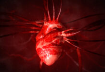
“Leonardo has long been recognized as one of the great artists of the Renaissance, but he was also a pioneer in understanding human anatomy. As an engineer and artist I wanted to know not only how the body was built, but also how it worked, ”says Declan O’Regan, head of the computational cardiac imaging group at the MRC Institute of Medical Sciences in London (UK) at Imperial College. In the early 16th century, Da Vinci first drew the intricate network of muscle fibers in the heart, called trabeculae, that form geometric patterns on the inner surface of the heart. Even then, the genius speculated that this network of fibers served to heat the blood as it flowed through the heart, but he had no way of verifying it.
Today, a team of scientists including O’Regan is closer to finding out. “Only now, with modern imaging techniques, computer simulations and genetics, can we begin to answer these questions,” says the cardiologist, whose research, published in the journal Nature , has relied on artificial intelligence to process 25,000 MRI images. of the heart, to which the associated genetic data were added. With all this information, the authors have concluded that the rough surface of the heart’s ventricles – its lower chambers – allows blood to flow more efficiently with each beat. They have also found that there are six regions in human DNA that would affect the development of fractal patterns in trabeculae.
Another conclusion of the study is that the shape of these muscle fibers influences the functioning of the heart, which could be related to heart disease. To confirm this, the scientists analyzed genetic data from 50,000 patients and found that different fractal patterns in this network had to do with the risk of developing heart failure – which occurs when the heart does not pump blood as it should. “We still need to understand how the blood that flows around the heart interacts with these muscle threads. This will help us identify patients at higher risk of developing heart disease complications, ”says O’Regan.
The cardiologist remembers that this is only one of the many questions that we remain unanswered about this powerful organ. To solve them, scientists are using techniques such as artificial intelligence, or very precise images such as magnetic resonance imaging, which allow them to easily dissect the human heart in 3D that even Leonardo could not have imagined.
These advances also include the bioartificial heart, which, to this day, remains a utopia. “Despite being a field with some years of experience, it still needs more research to become a reality, which I have no doubt we will see in the future”, maintains Carolina Gálvez-Montón, deputy director of the research group ICREC, which belongs to the Heart Institute of the Germans Trias i Pujol University Hospital (Barcelona). The studies are being carried out in vitro and with animal models, and consist of treating this organ as if it were new, removing all the animal cells but conserving its structures to repopulate them with a new muscular and vascular tissue compatible with that of the human being. This heart would be the one that would be transplanted to the person who needed it and would avoid having to depend on donated human organs. To reach this goal, scientists are working on several fronts: stem cells –with the potential to generate new tissues–, 3D prints –which are increasingly used to generate mini-organs– and computer simulation –to analyze contractions. and relaxations of the heart tissue of this new organ.
In Spain, Antoni Bayés-Genís, director of the Instituto del Corazón and the ICREC group, is one of the leaders. “The ICREC research laboratory has been investigating the artificial heart for more than a decade and two or three years ago it achieved a viable model for transfer to humans,” he highlights. Specifically, what they have developed is a sixteen-centimeter-square 3D structure that is made up of decellularized pericardium –the membrane that surrounds the heart– subsequently filled with a type of stem cells –MSC, derived from the umbilical cord. Scientists have started a phase 1 clinical trial with twelve patients. “In one or two years, we will be able to confirm its safety and advance to larger clinical trials to assess its efficacy,” says Bayés-Genís. The challenge of cardiac tissue engineering is enormous, but the possibilities it offers to improve the quality and quantity of life of our patients are also immense ”, adds the cardiologist.
Artificial tissue and organoids
While waiting for this biosanitary revolution to arrive, in parallel, scientists are developing new platforms to create viable artificial heart tissue and, also, cardiac organoids that mimic its functions and allow testing new drugs for different pathologies related to the heart. This, according to Gálvez-Montón, would avoid the use of animals in preclinical trials.
Regarding bioengineering to create tissues, Jane E. Freedman, director of cardiovascular research at the UMass Memorial Heart & Vascular Center (United States), confirms that test models are being developed, “ but we have not yet reached the point where we are clinically using blood vessels or bioengineering valves, although the research continues ”.
Minimally invasive surgery has also been a before and after for patients with heart problems. As the name suggests, these operations are characterized by the small incisions that the surgeon makes in the tissues in question, instead of having to open the patient’s chest. Unlike traditional surgeries, recovery is faster and allows a greater number of interventions in very old people, such as octogenarians or even nonagenarians.
“With minimally invasive techniques and transcatheter therapies, less aggressive procedures can be offered to the elderly patient with very good results”, comments Jorge Rodríguez-Roda, head of the Adult Cardiac Surgery Service at the Ramón y Cajal University Hospital (Madrid) and elected vice president of the Spanish Society of Cardiovascular and Endovascular Surgery (SECCE). The heart surgeon recalls that, a few years ago, common surgeries like these were unthinkable, especially those that refer to the implantation or repair of heart valves.
hand robot Da Vinci
Another advance is robotic surgery, with the Da Vinci robot as the maximum exponent. This device sits on the operating table like just another surgeon and helps real doctors operate on hearts, such as repairing mitral valves – which separate the two chambers on the left side of the heart – instead of having to replace them with prostheses. However, “its use is still very scarce due to its high cost and complexity in cardiac surgery,” argues Rodríguez-Roda.
Along the same lines, artificial intelligence techniques are also greatly helping cardiologists, who use them on a daily basis. As we have seen, one example is how they helped process the 25,000 MRI images of the heart to find out what is the function of the network of muscle fibers that intrigued the genius of the Renaissance.
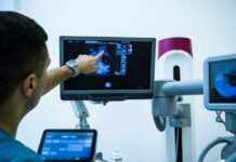
“In cardiac imaging, multiple machines – computed tomography, magnetic resonance imaging or echocardiography – use these technologies, which increases diagnostic capacity and saves time for specialists, by automatically performing multiple measurements using artificial intelligence algorithms,” he details. Dr. Rafael Vidal, member of the Executive Committee of the Spanish Society of Cardiology (SEC). According to the cardiologist, the future looks promising, since traditional tools such as the electrocardiogram, when digitized, will also benefit from artificial intelligence. ” It will make it possible to accurately predict if the heart has lost strength or if it is going to develop a serious arrhythmia in the future, which will help to establish preventive treatments or to start the search for the underlying pathology early ,” he explains.
Cardiologist Freedman agrees with him, who recalls that these techniques are already being used to obtain more information about the heart rhythm of patients, which can help doctors to find out who can suffer an arrhythmia and what type.
Massive genetic sequencing
In this journey through cardiac innovations, we cannot forget the contributions of the American Heart Association, a benchmark in world cardiology, which has been selecting the main advances in this field since 1996. Among them, massive genetic sequencing stands out. “It’s a fast-moving area,” Freedman declares. In addition to trying to answer Da Vinci’s questions, human genome studies have also provided cardiologists with more data on heart problems such as venous thromboembolism, which occurs when a blood clot forms in a deep vein, usually in the lower leg, thigh, or pelvis. If this clot breaks loose and travels through the bloodstream to the lungs, we speak of a pulmonary embolism, which can be fatal. A recent study published in the journal Nature Genetics that examined the DNA of more than 650,000 people found 22 new regions of the genome that correspond to this problem.
Another condition that can be serious is peripheral arterial disease, which occurs when the blood vessels outside the heart narrow due to the accumulation of fat and cholesterol in them. This can reduce or interrupt blood flow, usually to the legs, which can lead to tissue death and even foot or leg amputation. Research in Nature Medicine in which scientists analyzed the genetic information of more than 637,000 people identified eighteen new regions of the genome associated with this disease.
“This technology has radically changed the way we understand some cardiovascular diseases,” says Pablo García Pavía, from the Heart Failure and Family Heart Disease Unit of the Cardiology Service of the Puerta de Hierro University Hospital (Madrid). The cardiologist highlights that thanks to massive sequencing it has been possible to clarify the genetic cause of many hereditary heart diseases, such as cardiomyopathies, aortic diseases, cholesterol disorders and some cardiac arrhythmias, all of them associated with a high risk of death sudden.
“The knowledge we have of the genetic factors associated with an increased risk of very common diseases such as ischemic heart disease, high blood pressure and some metabolic diseases has also increased,” the specialist describes. Once they know the genes and mutations responsible for heart disease, cardiologists, thanks to massive DNA sequencing, can make the genetic diagnosis of their patients. “Establishing the genetic cause allows us to better estimate the risk and often establish the most appropriate treatment,” he says.
Precision medicine
This is what is known as personalized precision medicine, one that is tailored and adapted to each patient. According to García Pavía, thanks to genetic information, doctors can anticipate the disease and diagnose family members who are at risk of suffering from these pathologies before they manifest themselves. “Most likely, the new generations of cardiologists will use it as one more tool to give early diagnoses, and they will be able to establish cardiovascular prevention programs,” says specialist Gálvez-Montón.
And not only that. Mass sequencing has also been essential to develop specific treatments. ” Knowing new causal genes and new metabolic pathways has made it possible to develop new generation cardiovascular drugs “, recalls García Pavía. An example are some that treat very high cholesterol levels or those that act on DNA or messenger RNA.
Traditionally, for people at high risk of clots in their blood vessels, cardiologists have prescribed anticoagulants such as acenocoumarol, which needs constant monitoring, something that could go down in history. “One of the most important advances is the progressive replacement of vitamin K antagonists, which in Spain is the well-known Sintrom, by direct anticoagulants, which not only have the advantage of not requiring periodic blood tests, but also present less risk of complications ”, highlights Raúl Moreno, president of the SEC’s Interventional Cardiology Association.
In addition, the cardiologist points out that patients with severe hypercholesterolemia – with very high levels of cholesterol in the blood – are already receiving monoclonal antibodies subcutaneously once or twice a month. This new treatment reduces cholesterol levels when there is not a good response to the usual drugs, according to Moreno. In pharmacological treatment, new drugs aimed at patients with heart failure also stand out –sacubitril / valsartan–, by significantly reducing mortality and rehospitalizations, ”says Gálvez-Montón.
Seeing what has been achieved in recent years, scientists are optimistic. The bioartificial heart will be a reality although it is not easy to estimate a date. Tissue generation, cell therapies and minimally invasive surgeries will also advance more and more. Rodríguez-Roda also mentions xenografts, that is, heart transplants from animals, “although the risk of zoonoses makes their development very difficult,” he warns.
And it is expected that cooperation between the different specialties will increase in order to consolidate the combined treatments. If Leonardo da Vinci brought together an anatomist, an engineer and an artist in the same person, the collaboration between medical disciplines must follow the same philosophy: all together.